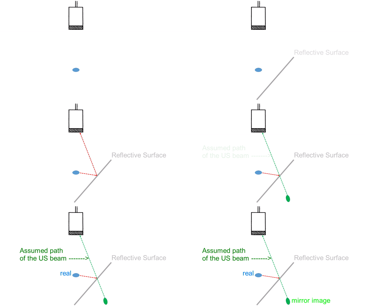
Mirror Artifact
All imaging artifacts results from violations of assumptions built into the machine’s software.
One of these assumptions is that US beams travel only in straight line, both from the probe to the imaged object and back from the object to the transducer. If a highly reflective, surface is present in the imaging field, some of the US beams may be reflected off that surface on the way from the transducer to the object and back. This “confuses” the machine and results in mapping of an object in a location beyond the reflective surface,where it actually does not exist.

Coronal view at posterior axillary line of costodiaphragmatic recess.
This probablyis the most commonly encountered example of mirror artifact. The lung is on the left side of the screen, and the liver – on the right. The two are separated by the diaphragm – a highly reflective surface.
Note that the spine “disappears” behind the lung, as it normally does, indicating that there is actually no consolidation here.
This is likely the second most common instance of mirror artifact. In this PLAx view a small pericardial effusion is seen posterior to the heart. Dense pericardium acts as a reflector and a second moving epicardial heart surface is seen where left lung should be.
Similarly, in this A4 view there is a clear mirror artifact accounting for the second lateral tricuspid annulus moving below the diaphragm.
Finally, a very unusual but spectacular example of mirror artifact.This 41 y.o. woman with ESRD and h/o severe hypercalcemia, has calcifications in multiple tissues, including her pleura and lung fissures. Here pleural effusion and atelectatic lung are reflected in the “mirror” of a densely calcified horizontal fissure.