Cornell
HM-POCUS
Hospital Medicine Point of Care Ultrasonography
Search
Login
Home
About Us
What is HM-POCUS?
Our Faculty
Contact Us
Testimonials
Education
5-Day POCUS Class
Registration
SHM/CHEST Certification
Itinerary
For Instructors
Instructions
Stations
Practical Exams
Optimization
Homework Videos
Case Conference
Remote Access
Case Conference Schedule
Case Conference Archive
Case Conference Archive: Cardiogenic Shock
Case Conference Archive: Cardiac Tamponade
Presentation Standards
[Column]
Resident-Elective
IM Fellows: POCUS Course
Medical Students
Interns
Faculty
PAs
Practice Questions
Ultrasound Machines
Our Ultrasound Machines
Technical Documentation
Specs
Quick Card
Transducers
User Guide
Basic User Manual
Basic Service Manual
Advanced Reference Manual
[Column]
Machine Storage
Machine Access
Cleaning the Machines
Advanced Cardiac Ultrasound
Missing Ultrasound Machine
Credentialing
Portfolio
Privileges and Certification
Procedures
Consent Forms
Consent for CVL
Consent for HD Catheter
Consent for LP
Consent for Paracentesis
Consent for Thoracentesis
DIY, Videos, Etc
LP Technique
Spine Anatomy
Paracentesis Technique
Thoracentesis Technique
[Column]
Moderate Sedation
Sterile Technique
Using Peripheral Venous Caths Instead of Standard Kits
Getting Supplies
Procedure Log
Suturing Supplies
Saving Images
Image Formatting
Saving Images to a Hard Drive
Hard Drive to Q-Path
Hard Drive to USB
Resources
Menu
Home
About Us
What is HM-POCUS?
Our Faculty
Contact Us
Testimonials
Education
5-Day POCUS Class
Registration
SHM/CHEST Certification
Itinerary
For Instructors
Instructions
Stations
Practical Exams
Optimization
Homework Videos
Case Conference
Remote Access
Case Conference Schedule
Case Conference Archive
Case Conference Archive: Cardiogenic Shock
Case Conference Archive: Cardiac Tamponade
Presentation Standards
[Column]
Resident-Elective
IM Fellows: POCUS Course
Medical Students
Interns
Faculty
PAs
Practice Questions
Ultrasound Machines
Our Ultrasound Machines
Technical Documentation
Specs
Quick Card
Transducers
User Guide
Basic User Manual
Basic Service Manual
Advanced Reference Manual
[Column]
Machine Storage
Machine Access
Cleaning the Machines
Advanced Cardiac Ultrasound
Missing Ultrasound Machine
Credentialing
Portfolio
Privileges and Certification
Procedures
Consent Forms
Consent for CVL
Consent for HD Catheter
Consent for LP
Consent for Paracentesis
Consent for Thoracentesis
DIY, Videos, Etc
LP Technique
Spine Anatomy
Paracentesis Technique
Thoracentesis Technique
[Column]
Moderate Sedation
Sterile Technique
Using Peripheral Venous Caths Instead of Standard Kits
Getting Supplies
Procedure Log
Suturing Supplies
Saving Images
Image Formatting
Saving Images to a Hard Drive
Hard Drive to Q-Path
Hard Drive to USB
Resources
Cornell HM-POCUS
Menu
Home
About Us
What is HM-POCUS?
Our Faculty
Contact Us
Testimonials
Education
5-Day POCUS Class
Registration
SHM/CHEST Certification
Itinerary
For Instructors
Instructions
Stations
Practical Exams
Optimization
Homework Videos
Case Conference
Remote Access
Case Conference Schedule
Case Conference Archive
Case Conference Archive: Cardiogenic Shock
Case Conference Archive: Cardiac Tamponade
Presentation Standards
[Column]
Resident-Elective
IM Fellows: POCUS Course
Medical Students
Interns
Faculty
PAs
Practice Questions
Ultrasound Machines
Our Ultrasound Machines
Technical Documentation
Specs
Quick Card
Transducers
User Guide
Basic User Manual
Basic Service Manual
Advanced Reference Manual
[Column]
Machine Storage
Machine Access
Cleaning the Machines
Advanced Cardiac Ultrasound
Missing Ultrasound Machine
Credentialing
Portfolio
Privileges and Certification
Procedures
Consent Forms
Consent for CVL
Consent for HD Catheter
Consent for LP
Consent for Paracentesis
Consent for Thoracentesis
DIY, Videos, Etc
LP Technique
Spine Anatomy
Paracentesis Technique
Thoracentesis Technique
[Column]
Moderate Sedation
Sterile Technique
Using Peripheral Venous Caths Instead of Standard Kits
Getting Supplies
Procedure Log
Suturing Supplies
Saving Images
Image Formatting
Saving Images to a Hard Drive
Hard Drive to Q-Path
Hard Drive to USB
Resources
Login
HM-POCUS for Faculty
HM-POCUS for Faculty Class 1
Requirements
Itinerary
Practical Skills Exam
Reading List
Curriculum
Curriculum 1
Mirror Artifact
Curriculum 1: Renal and Bladder
Curriculum 1: Lungs and Pleura
Hemothorax
Pneumothorax
Consolidation
Anterior Consolidation
Curriculum 1: Lower Extremity DVT
Curriculum 1: Basic Cardiac Ultrasound
Advanced Cardiac Ultrasound
Doppler
Advanced Cardiac Views
Diastology
Valvular DZ
PA Pressure
RV 2
Hemodynamics
Forgotten Structures
Coronary Sinus
LA Appendage
Pulmonary Veins
Curriculum 1: US Basics
US Safety
Curriculum 1: Abdominal Aorta
Curriculum 2
Curriculum 2: Optic Nerve
Curriculum 2: Biliary
Curriculum 2: Musculoskeletal US
Curriculum 2: Heart
Advanced Cardiac Ultrasound
Doppler
Advanced Cardiac Views
Diastology
Valvular DZ
PA Pressure
RV 2
Hemodynamics
Forgotten Structures
Coronary Sinus
LA Appendage
Pulmonary Veins
Practice Questions
Image Review
Menu
HM-POCUS for Faculty Class 1
Requirements
Itinerary
Practical Skills Exam
Reading List
Curriculum
Curriculum 1
Mirror Artifact
Curriculum 1: Renal and Bladder
Curriculum 1: Lungs and Pleura
Hemothorax
Pneumothorax
Consolidation
Anterior Consolidation
Curriculum 1: Lower Extremity DVT
Curriculum 1: Basic Cardiac Ultrasound
Advanced Cardiac Ultrasound
Doppler
Advanced Cardiac Views
Diastology
Valvular DZ
PA Pressure
RV 2
Hemodynamics
Forgotten Structures
Coronary Sinus
LA Appendage
Pulmonary Veins
Curriculum 1: US Basics
US Safety
Curriculum 1: Abdominal Aorta
Curriculum 2
Curriculum 2: Optic Nerve
Curriculum 2: Biliary
Curriculum 2: Musculoskeletal US
Curriculum 2: Heart
Advanced Cardiac Ultrasound
Doppler
Advanced Cardiac Views
Diastology
Valvular DZ
PA Pressure
RV 2
Hemodynamics
Forgotten Structures
Coronary Sinus
LA Appendage
Pulmonary Veins
Practice Questions
Image Review
Basic Cardiac Ultrasound
Sections
Basic Views
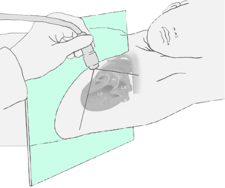
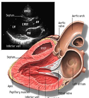
PLAx
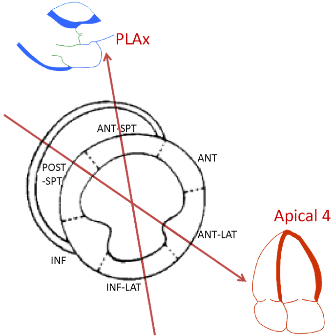
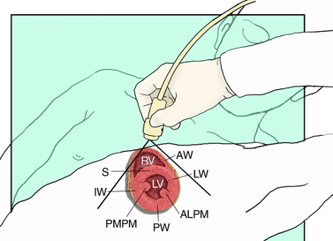
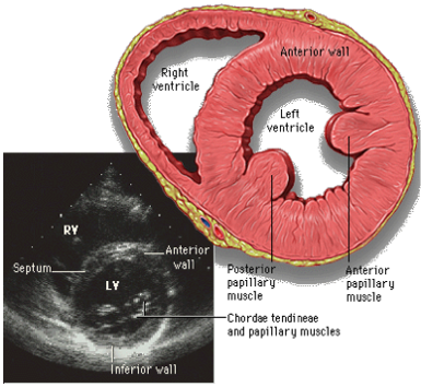
PSAx
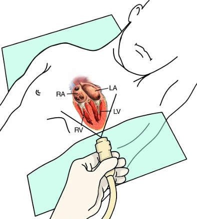
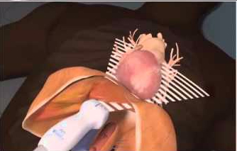
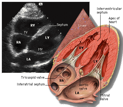
Apical 4
Subcostal
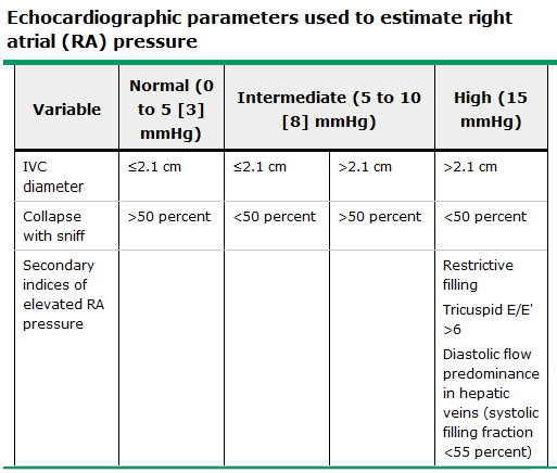
IVC
LV Systolic Fx
Intro to Doppler
M-mode
RV
Tamponade
Basic Views
Parasternal Long Axis (PLAx)
Parasternal Short Axis (PSAx)
Apical 4-Chamber View (A4)
Subxiphoid Long-Axis
Subcostal IVC View
LV Systolic Function
Introduction to Doppler
Qualitative Assessment of
Severity of Regurgitation
by Ratio of Area of the Jet to the Area of Receiving Chamber
< 20% – mild
25-30% – moderate
30-40% – moderate to severe
>40% – severe
M-Mode
The Right Ventricle
RV Size
RV Function
Tamponade
Login
Log In
Lost your password?