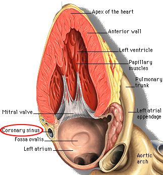
A main vein collecting blood from the heart muscle and draining into the RA.
Who cares about Coronary Sinus?
Coronary sinus (CS) is an important landmark, and knowledge of normal anatomy will facilitate identification of structures and pathology.
Movement of CS during the cardiac cycle corresponds to the movement of mitral annuls and thus may be useful in assessing LV systolic function (formalized as MAPSE) (ref).
Dilated CS is seen in cases of elevated RA pressure, or in congenital anomalies resulting in increased blood flow through the CS (such as persistence of left IVC). Of these, of course, acquired causes, such as RV failure (ref, ref), severe TR and pulmonary HTN are most important for an internist.
Coronary Sinus on TTE
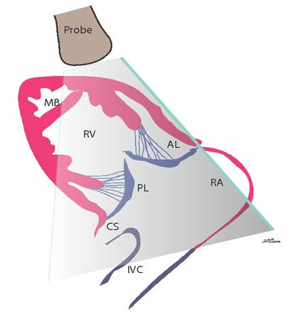
Coronary sinus is located in the left AV groove, between the left ventricle and the left atrium. CS can be visualized in the following windows:
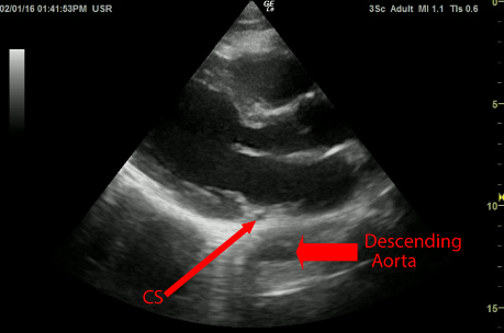
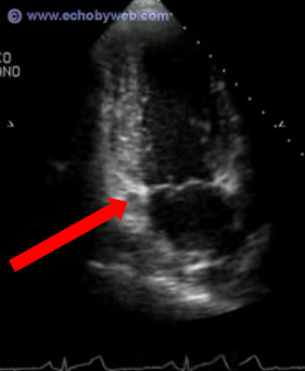
Normally in PLAx view coronary sinus appears as a small unechoic dot between LA and the LV. It is located within pericardial sack, unlike descending aorta. In normal individuals the two cannot really be confused. Note howCS moves along the long axis of the heart during the cardiac cycle.
Note severely decreased excursion of the CS during the cardiac cycle, corresponding to severely decreased LV systolic fx.
This is clip shows incidentally discovered dilated CS. The patient was confirmed to have persistent Left SVC. Descending aorta can be seen outside the heart.
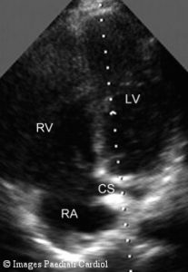
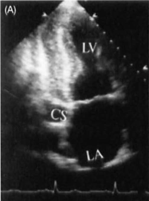
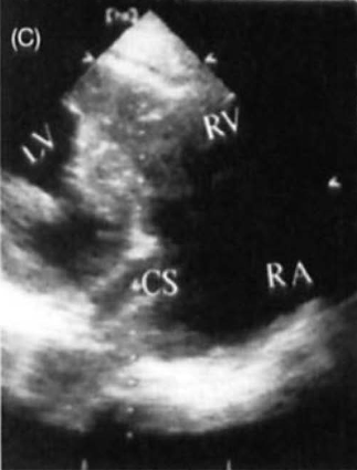
Apical 4-Chamber, Coronary Sinus View (A4-CS)
CS is a posterior cardiac structure, and in order to visualize it from the apex, one has to tilt the probe posteriorly (dorsally) from the usual A4 position. Sometimes inadvertently this view is obtained instead of a proper A4.
Normal CS
Note that normal CS is a curved structure which can rarely be imaged along its entire course. Most commonly, only the most distal portion of CS is imaged, including its opening into the RA. In the images below the normal CS appears as a tubular structure between LA and LV.
Quilina, O., et al. Normal adult echocardiography – apical views. Images in Paediatric Cardiology. 2007;9(1):1-9.
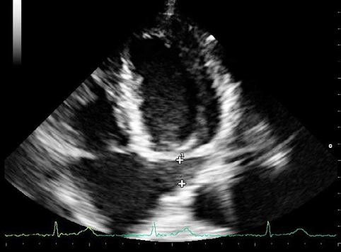
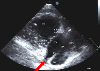
Dilated CS
Because scanning plane in A4-CS view is usually oblique to the axis of CS, the sinus frequently appears not as a tube, but as a horizontal cone, with its base at the opening into the RA. In all this cases one is looking behind the LA for the CS.